Seeing the invisible
Dr. Chung-Min Kang explains how quantitative light-induced fluorescence (QLF) technology promotes minimally invasive diagnostics, trust, and understanding between pediatric dentists and patients.
Dr. Chung-Min Kang discusses the transformative role of quantitative light-induced fluorescence technology in pediatric caries diagnosis
In pediatric dentistry, early detection is everything. Yet, for clinicians working with very young or behaviorally uncooperative children, conventional tools often fall short. Enter quantitative light-induced fluorescence (QLF) technology, an innovative technique in non-invasive caries detection that is gaining traction across Korean clinics — and increasingly, across the global dental landscape.
What is QLF?
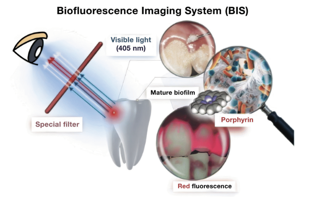
Quantitative light-induced fluorescence allows clinicians to visualize dental lesions by detecting fluorescence loss in demineralized enamel. Its key strength lies in capturing early caries that may not yet appear on radiographs, and doing so without radiation exposure. For pediatric practitioners, this means faster, more comfortable exams — and fewer tears in the chair.
A device that provides this type of fluorescence, called Qray, provides quantitative data such as the ∆F value (fluorescence loss percentage) and lesion area, which can be used to track progression or arrest over time. A greater ∆F value has been associated with active lesions requiring clinical attention, while smaller fluorescence losses may indicate arrested or remineralized lesions.
Why use this technology for children?
Children present unique diagnostic challenges. Cooperation may be limited, lesion progression is often rapid, and parental involvement is critical. This technology offers a real-time, visual, and objective caries detection method, especially valuable in:
- Early caries detection in anterior and posterior teeth
- Behavioral management, allowing for quick, non-invasive assessments
- Parental communication, with fluorescence images making caries visibly evident, enhancing understanding and treatment acceptance
- Monitoring lesion progression or arrest over time
- Reducing radiation exposure, aligning with ALARA principles
In Korea, this technology has received new medical technology accreditation and is currently covered under the country’s national health insurance, reflecting strong clinical support and public health value.
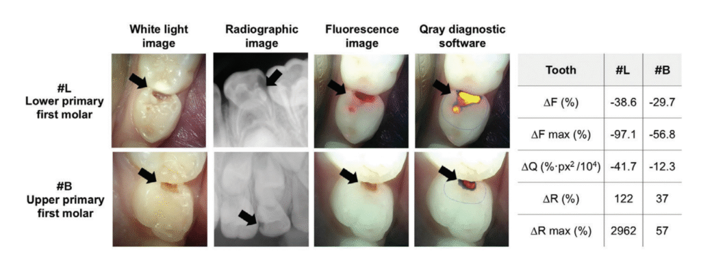
Although the visual size of the lesions appears similar, analysis with this device shows significantly higher quantitative values in tooth #L (∆F: −38.6%, ∆Q: −41.7, ∆R: 122%) compared to tooth #B. This indicates that the lesion in #L is more active and likely to progress, demonstrating the value of this device in objectively assessing lesion severity beyond visual inspection.
What the research says
Multiple studies have validated Qray’s diagnostic efficacy in primary teeth. In a study by Cho, et al. (2021), Qray demonstrated 88% sensitivity and 90% specificity for detecting dentin caries in primary molars compared with histologic validation.1 A 2022 in vitro study confirmed its robustness in controlled conditions, showing clear separation between sound and carious tissues based on ∆F thresholds.2
Qray also enables clinicians to evaluate caries activity. Kim, et al. (2022), found that active lesions exhibited significantly larger ∆F values and lesion areas, and were associated with distinct microbial profiles, including higher levels of Streptococcus mutans and Lactobacillus spp.3 This opens the door for a more biologically informed approach to caries management, reducing overtreatment of inactive lesions.
In a 2023 randomized controlled clinical trial, Qray was used to assess lesion depth and healing in pulpotomized primary molars, showing potential as a non-invasive follow-up tool for restorative outcome evaluation.4
The Future is bright
As dentistry continues to embrace minimally invasive diagnostics and technology-assisted care, quantitative light-induced fluorescence (QLF) technology is well-positioned as a bridge between clinical accuracy and child-friendly care. For providers in pediatric and preventive settings, this tool doesn’t just illuminate teeth — it illuminates trust, understanding, and better outcomes.
 Chung-Min Kang, DDS, PhD, is an Associate Professor, at the Department of Pediatric Dentistry, Yonsei University College of Dentistry, Seoul, South Korea. Disclosure: Dr. Kang reports no financial conflicts of interest.
- Cho KH, Kang CM, Jung HI, Lee HS, Lee K, Lee TY, Song JS. The diagnostic efficacy of quantitative light-induced fluorescence in detection of dental caries of primary teeth. J Dent. 2021 Dec;115:103845. doi: 10.1016/j.jdent.2021.103845. Epub 2021 Oct 9.
- Cho KH, Kang CM, Jung HI, Lee TY, Song JS. Assessment of the caries detection ability of quantitative light-induced fluorescence (QLF) in primary teeth in vitro. Journal Korean Acad Pediatr Dent. 2022;49(1):65-75. doi: http://doi.org/10.5933/JKAPD.2022.49.1.65.
- Kim CH, Bae K, Lee TY, Song JS, Kim SO, Kang CM. Assessment of dental caries lesion activity status using quantitative parameters obtained from the quantitative light-induced fluorescence method and difference of microbial distribution in primary molars. Photodiagnosis Photodyn Ther. 2022 Sep;39:102942. doi: 10.1016/j.pdpdt.2022.102942. Epub 2022 May 28.
- Joo Y, Lee T, Jeong SJ, Lee JH, Song JS, Kang CM. A randomized controlled clinical trial of premixed calcium silicate-based cements for pulpotomy in primary molars. J Dent. 2023 Oct;137:104684. doi: 10.1016/j.jdent.2023.104684. Epub 2023 Sep 1.